Location
710e (Shenzhen Research Institute)
Ms. Dexiu ZHENG
Assistant Scientific officer
- 611b (Shenzhen Research Institute)
- Dexiu.zheng@polyu.edu.hk
Equipment and Service
ULS-SZ
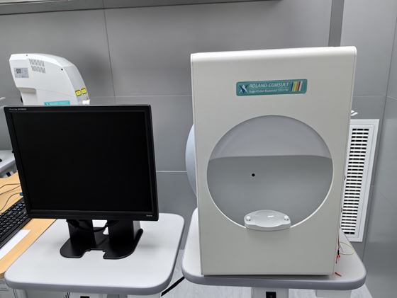
Electrophysiological Test Unit is usable for Pattern VEP+ Pattern ERG + Flash VEP, for scotopic and photopic ERG, EOG fast and slow, mf ERG Flash stimulation and mf VEP Pattern stimulation. The Roland RETI-Scan animal unit consists of the stimulator units and data recording and analyzing system. The biosignal amplifier includes a preamplifier near the patient. All patient data and the results are stored in a database. The biosignal and averaged curves from all channels can displayed on the monitor. In the analyze mode the system set all markers and calculates all defined parameters automatically. The software includes a lot of advanced features like FFT analyze, off-line averaging and PreTrigger. The new Macula tool for better and accurate understanding of function and structure of the Retina. It is possible to do in 30deg SLO picture, ERG for 7 and 19 areas.
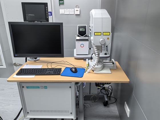
The product is comprised of multiple components, including a stimulator which encompasses a graphic stimulator and a flash stimulator. Other essential elements consist of a signal amplifier, an animal experimental table equipped with a flexible electrode holder, a thermostat for temperature regulation, and a variety of electrodes such as animal needle electrodes, animal ring cornea electrodes of varying sizes (4mm, 8mm, and 3mm), ERG contact lens electrode, and DTL electrode. Additional items include DTL connection wire, mouse corneal contact lens, mouse lens, flash stimulator, computer, display, keyboard, mouse, power box, and electrode.
710e (Shenzhen Research Institute)
Assistant Scientific officer
We use Cookies to give you a better experience on our website. By continuing to browse the site without changing your privacy settings, you are consenting to our use of Cookies. For more information, please see our Privacy Policy Statement.
Your browser is not the latest version. If you continue to browse our website, Some pages may not function properly.
You are recommended to upgrade to a newer version or switch to a different browser. A list of the web browsers that we support can be found here