Sound Solution: A Breakthrough Approach to Parkinson's Disease
Other Articles

Harnessing Hollow Silica Nanostructures for Precise, Durable and Clinically Relevant Ultrasound Brain Stimulation
Study conducted by Prof. Lei SUN and his research team
Parkinson's disease (PD) is one of the most common neurological disorders associated with later life, and its prevalence rises sharply with age. It affects the brain systems that control movement, often causing tremor, stiffness, slowness and problems with balance, all of which can reduce independence and quality of life in older adults. The condition is mainly linked to the gradual loss of dopamine-producing nerve cells. Current treatment usually relies on medicines such as levodopa, which can improve symptoms, especially in the earlier stages, but their benefit may become less consistent over time. For some patients, deep brain stimulation (DBS) offers another option, yet this involves surgery to place electrodes deep inside the brain.
These limitations continue to motivate the search for neuromodulation approaches that are both precise and less invasive. In a recent study published in Nature Communications [1], Prof. Lei SUN, Professor of the Department of Biomedical Engineering at The Hong Kong Polytechnic University, and his research team address the need for such approaches by combining low-intensity ultrasound with engineered hollow silica nanostructures (HSN) to achieve chronic, localised brain stimulation in mice, as well as therapeutic benefit in PD models.
The principal advancement of this work is the demonstration that ultrasound neuromodulation can be made both spatially precise and durable through the use of stable, sonoresponsive nanostructures. Conventional transcranial ultrasound is attractive because it penetrates deeply and can be delivered non-invasively, but its spatial resolution is typically limited to millimetre or centimetre scales, making selective activation of small neuronal populations difficult. Earlier nanoparticle- or nanobubble-assisted approaches improved localisation, but their in vivo instability limited usefulness for chronic applications. In contrast, the HSN developed by Prof. Sun and his team persisted in the mouse brain for more than nine weeks while remaining functionally responsive to ultrasound. This enabled repeated stimulation of targeted regions, including the motor cortex, striatum, ventral tegmental area (VTA) and subthalamic nucleus (STN), without genetic modification and without repeated intracranial administration.
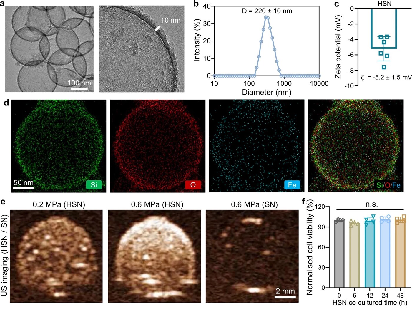
Figure 1. Characterisation of the fabricated HSN
(a) Transmission electron microscopy image of HSN
(b) Size distribution plot of HSN
(c) Zeta potential measurement of HSN
(d) EDS elemental mappings of HSN
(e) Ultrasound imaging of HSN and SN under varying acoustic energy levels
(f) Cytotoxicity assessment of HSN
The HSN were designed as hollow, gas-containing silica particles with an approximately 10 nm shell and an overall diameter of around 220 nm. The characterisation of the HSN is illustrated in Figure 1. The hollow architecture is central to their function, as it allows them to amplify local acoustic effects under ultrasound exposure. Iron was incorporated into the shell to improve robustness and to provide magnetic responsiveness, creating the possibility of MRI visibility. To improve colloidal stability and biocompatibility, the surface was modified with polyethylene glycol (PEG).
Characterisation confirmed a spherical morphology, uniform elemental distribution of silicon, oxygen and iron (Figure 1d), and successful PEG functionalisation. The particles produced strong ultrasound contrast signals, unlike solid silica nanoparticles (SN) used as controls (Figure 1e), confirming that the gas-filled hollow structure was responsible for their acoustic responsiveness. They also retained contrast under repeated ultrasound bursts, indicating substantial mechanical stability. Importantly, the particles showed no detectable cytotoxicity in primary neuronal cultures (Figure 1f).
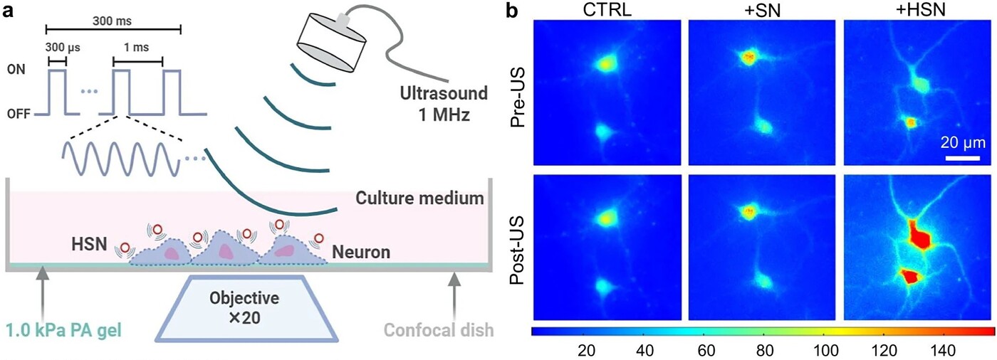
Figure 2. HSN-enabled low-intensity ultrasound modulation of primary neurons
(a) Schematic illustration depicting HSN-medicated ultrasound stimulation of primary cultured neurons
(b) Representative images showing calcium fluorescence in primary neurons without nanoparticles (CTRL) or with HSN/SN, before and after ultrasound stimulations
The study then examined whether HSN could enhance neuronal responsiveness to low-intensity ultrasound. Primary cortical neurons were grown on an acoustically transparent polyacrylamide (PA) gel and then exposed to ultrasound sonication (Figure 2a). Calcium responses were visually recorded. In cultured primary cortical neurons, ultrasound in the presence of HSN produced marked calcium influx, whereas ultrasound alone (CTRL) or ultrasound with SN did not (Figure 2b).
Responses were repeatable, rapidly reversible and dependent on acoustic intensity. The team further tested the mechanism using gadolinium, a broad inhibitor of mechanosensitive ion channels. This substantially suppressed the calcium signal, with recovery after washout, supporting the conclusion that HSN-mediated ultrasound acts primarily through activation of mechanosensitive ion channels. This interpretation was reinforced by increased c-Fos expression, indicating downstream neuronal activation. Together, these data show that HSN sensitise nearby neurons to mechanical stimulation by ultrasound, allowing effective activation at relatively low acoustic intensities.
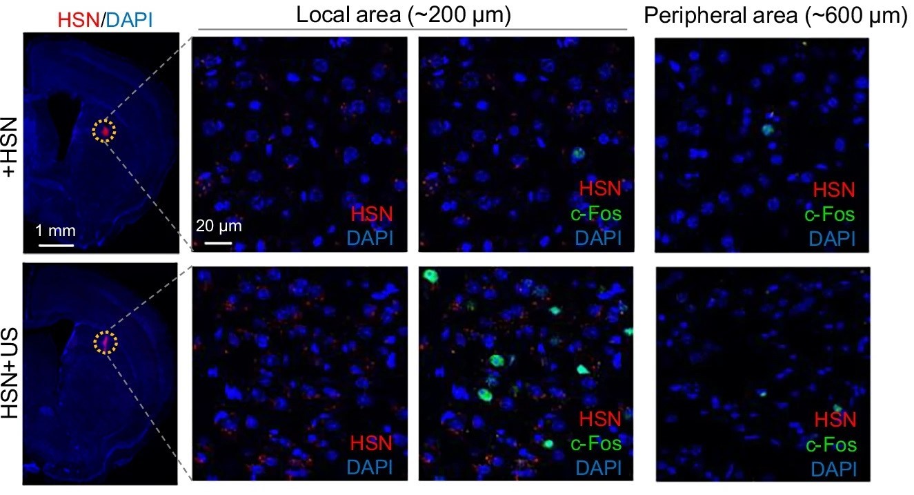
Figure 3. Immunofluorescence staining of c-Fos expression (green) in the area surrounding the HSN (red) injection site in the mouse stratum stimulated with or without US
In vivo findings were consistent with the in vitro data and demonstrated a high degree of localisation. After HSN injection into the motor cortex of a mouse, low-intensity ultrasound elicited forelimb electromyographic responses, while saline controls showed little or no response at lower pressures. Response magnitude and success rate increased with HSN dose, indicating a controllable effect. In the striatum, c-Fos activation following ultrasound was confined to the HSN-containing region, with little increase in adjacent tissue lacking particles (Figure 3). Notably, the activated zone was substantially smaller than the ultrasound wavelength itself. This is an important conceptual point: spatial selectivity was determined not by focusing ultrasound more tightly, but by restricting where the sonosensitive nanostructures were located. The study therefore shifts precision from the acoustic field alone to the combined acoustic–material interface.
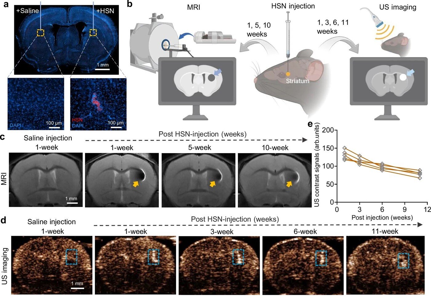
Figure 4. In vivo HSN lifetime test using fluorescence, MRI and ultrasound imaging.
(a) Representative fluorescence images of mouse brain slices following HSN and Saline injection at week 9
(b) Schematic illustration of the experimental process
(c) MRI images of mice brains at different time points after HSN/Saline injection
(d) Ultrasound contrast-mode images of mice brains at different time points after HSN/Saline injection
A major strength of the work is its demonstration of chronic utility. Fluorescence imaging, MRI and ultrasound imaging all showed persistence of HSN in the brain for over two months (Figure 4), with gradual signal decline consistent with biodegradation and clearance. In the VTA, fibre photometry using GCaMP6s showed that ultrasound continued to evoke rapid and reversible calcium responses up to nine weeks after HSN injection. Although response amplitude declined modestly over time, latency remained stable, indicating durable functional performance. Histological analysis suggested that extracellular persistence was accompanied by uptake into astrocytes, neurons and especially microglia, implicating microglia-mediated clearance as a likely pathway. For chronic neurological disorders, this balance between persistence and eventual biodegradation is highly relevant.
The most translationally important aspect of the study is its application to PD models. In an MPTP-induced mouse model, the team injected HSN into the STN, a clinically established DBS target, and delivered repeated ultrasound over nine weeks. Mice receiving HSN plus ultrasound showed progressive improvement in rotarod performance, indicating better motor coordination, while saline-treated controls did not. Open-field testing similarly showed increased locomotor activity and mobile time in the treated group. Importantly, these gains persisted for at least two weeks after the treatment period ended, suggesting more than an immediate stimulation effect.
To understand how the treatment worked, the team examined dopamine activity in brain circuits linked to movement. Ultrasound stimulation of the HSN-targeted STN increased dopamine signalling, suggesting that the approach activated pathways affected in Parkinson’s disease. Treated mice also showed better preservation of dopamine-related neurons. Similar benefits were seen in a second Parkinson’s model, indicating that this ultrasound-based method may achieve some DBS-like effects without implanted electrodes.
The safety results were encouraging. Over two months, mice given HSN maintained normal weight and no deaths were recorded. Tests of movement, memory and cognition did not show clear differences from control animals. Brain tissue analysis also found no obvious signs of cell death, inflammation or damage. Because the study used low-intensity ultrasound, the risk of overheating or physical injury was also reduced. Even so, longer-term safety studies will still be needed before clinical use.
In summary, this study provides a compelling proof of concept for chronic, localised and minimally invasive neuromodulation using ultrasound and hollow silica nanostructures. The work is particularly relevant to age-related neurological disease because it addresses a central translational challenge: how to stimulate deep brain circuits precisely, repeatedly and safely without implanted hardware or genetic manipulation. For PD and, potentially, other chronic neurological disorders, it suggests a future in which engineered nanomaterials and focused acoustic energy may complement or in some cases reduce reliance on, conventional invasive neuromodulation strategies.
Prof. Sun has 25 years of research and development experience in medical ultrasound technology, including in system development, signal/image processing and biomedical applications. He holds more than ten US and Chinese patents, either granted or filed. As a Principal Investigator, he has led research projects in Hong Kong and Chinese mainland with total funding of over HKD76 million. In recognition of Prof. Sun's outstanding research achievements in the field of biomedical engineering, he was awarded a fellowship under the Research Grants Council's (RGC) Senior Research Fellow Scheme (SRFS) 2025/26.
| References |
|---|
[1] Hou, X., Jing, J., Shi, Z., Jiang, Y. & Sun L. (2026). Sono-mechanical nanostructures-enabled sustained precise ultrasound brain stimulation. Nature Communications 17, 3060 (2026). https://doi.org/10.1038/s41467-026-69710-8
 | Prof. Lei SUN Professor, Department of Biomedical Engineering |