
Beyond BMI: How Fat Distribution Shapes the Ageing Brain
Other Articles

Why Regional Adiposity Matters for Cognition, Brain Health and Ageing
Study conducted by Prof. Anqi QIU and her research team
As populations age, protecting brain health has become a central priority for the smart ageing industry. Cognitive decline affects independence, care demand and quality of life, while also increasing the risk of neurodegenerative conditions such as Alzheimer's disease. This has intensified interest in modifiable midlife risk factors that may influence how the brain ages. Obesity is one such factor. However, research has often relied on body mass index (BMI) as the main measure. BMI is useful for estimating general obesity, yet it does not show where fat is stored. That is a major limitation, because fat stored in different parts of the body has different biological properties. Visceral fat, for example, is more strongly linked to inflammation and metabolic dysfunction than fat stored elsewhere. For brain ageing research, this means BMI captures only part of the picture.
A study published in Nature Mental Health [1] addresses this gap directly. Using UK Biobank data, Prof. Anqi QIU, Global STEM Scholar and Professor of the Department of Health Technology and Informatics at The Hong Kong Polytechnic University, and her research team proved that regional fat distribution is associated with brain structure, functional connectivity, white-matter integrity and cognition—independently of BMI. Fat stored in the arms, trunk, legs and visceral region showed distinct relationships with the brain, particularly in the sensorimotor, limbic, default mode and subcortical–cerebellar–brainstem systems.
The study also found that older-looking cortical brain systems mediated the relationship between adiposity and poorer cognition. Among all fat depots, visceral adiposity showed the strongest adverse associations with brain integrity and cognitive performance.
The study drew on a very large sample from the UK Biobank. Regional fat measures for arm, leg and trunk adiposity were available in over 23,000 adults with multimodal MRI data, while visceral adipose tissue data were available in nearly 19,000. Regional adiposity was measured by dual-energy X-ray absorptiometry, which provides a more precise estimate of body fat distribution than anthropometric measures. Brain health was assessed using structural MRI, resting-state functional MRI and diffusion imaging. Cognitive tests covered reasoning, executive function, processing speed and memory. This multimodal design allowed the team to examine the brain–fat relationship in far greater detail than studies using BMI alone.
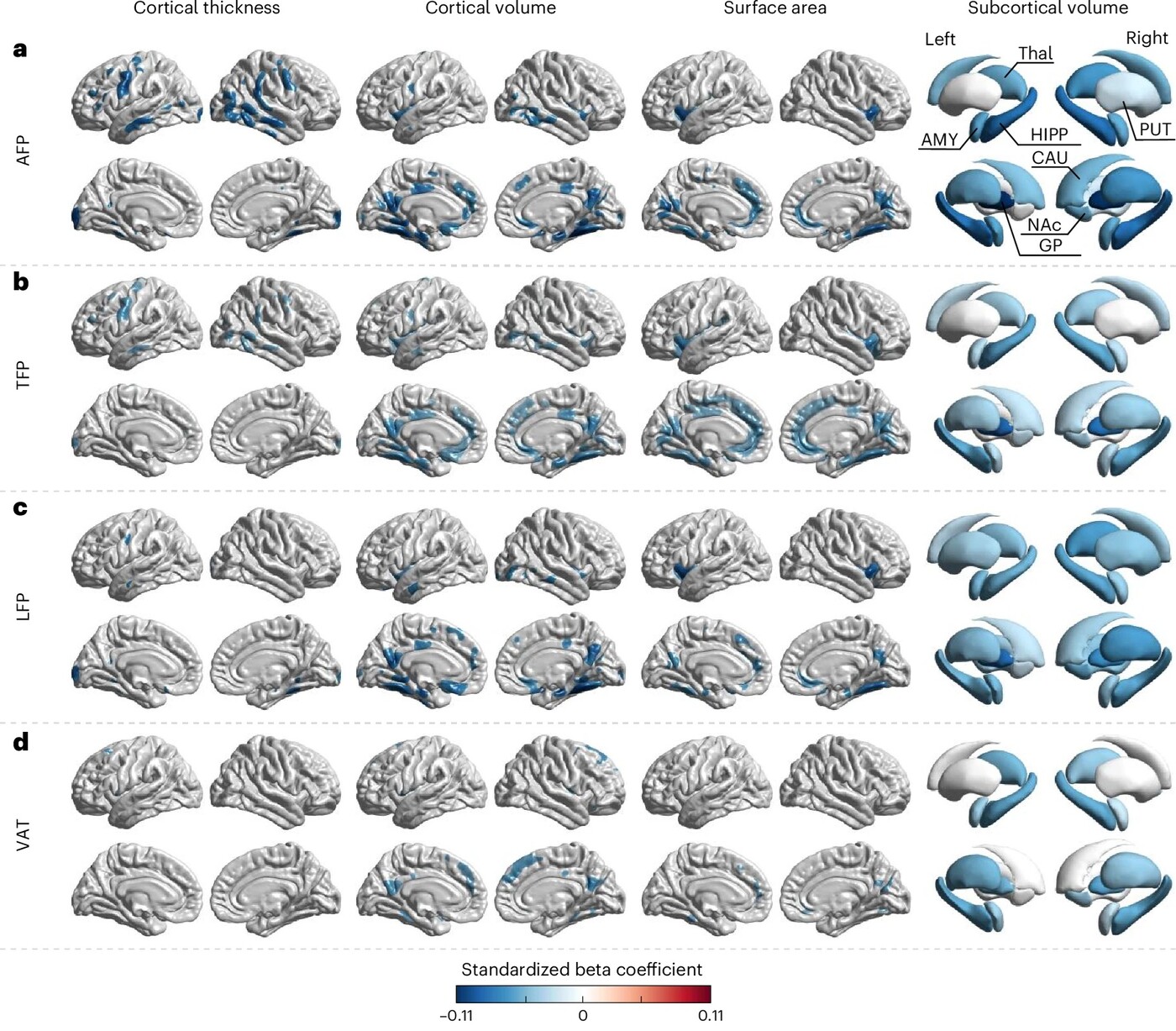
Figure 1. Associations of regional adiposity with cortical and subcortical morphometry. The blue colour indicates negative correlations. AFP: arm fat percentage; TFP: trunk fat percentage; LFP: leg fat percentage; VAT: visceral adipose tissue
The structural MRI results showed that regional adiposity was associated with selective cortical and subcortical changes. Higher arm, trunk and leg fat percentages were linked to reduced cortical volume and surface area in regions related to the default mode network (Figure 1), including the medial prefrontal cortex, posterior cingulate, precuneus and lateral temporal cortex. These regions are highly relevant to ageing and Alzheimer's disease, as the default mode network is frequently implicated in early neurodegeneration. The same adiposity measures were also associated with changes in limbic-related areas such as the medial temporal and orbitofrontal cortex. Arm and trunk fat were additionally linked to reduced cortical thickness in the sensorimotor cortex. Visceral adiposity showed a more focused pattern, with prominent associations in key default mode hubs such as the dorsal medial prefrontal cortex and precuneus. All four adiposity measures were negatively associated with subcortical volumes.
The functional connectivity findings revealed a partly overlapping but distinct pattern. All adiposity measures were associated with reduced connectivity in the sensorimotor network and in subcortical–cerebellar–brainstem circuits. These systems are important for motor control, autonomic regulation and core homeostatic processes. Leg fat showed the strongest negative relationship with connectivity in the limbic network, which is involved in emotion, memory and reward. Arm, leg and trunk adiposity were associated with increased within-network connectivity in the default mode network, whereas visceral fat was not significantly associated with default mode connectivity.
This mismatch between structural and functional findings suggests that fat-related brain changes may not occur uniformly across imaging modalities. Structural vulnerability may be detectable before clear connectivity disruption, or compensatory mechanisms may temporarily preserve network function.
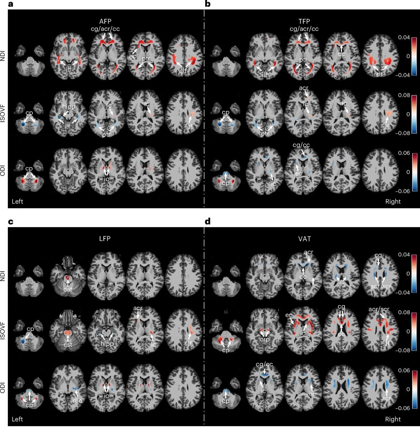
Figure 2. Association of fat distribution with neurite orientation dispersion and density imaging metrics. Regions with significant associations are highlighted. The red colour indicates positive correlations while the blue colour indicates negative correlations. NDI: neurite density index; ISOVF: isotropic volume fraction; ODI: orientation dispersion index
The white-matter findings (Figure 2) were especially important because they differentiated visceral fat from other depots more clearly. Visceral adiposity was associated with lower neurite density, higher free-water contamination and lower axonal organisation across widespread white-matter tracts which included the corona radiata, internal and external capsules, corpus callosum and cingulum. Taken together, these patterns point to compromised white-matter integrity and possible tissue degeneration.
By contrast, arm, trunk and leg adiposity showed more mixed and less consistently adverse associations with white matter. For ageing research, this is highly relevant because white-matter integrity supports efficient communication across neural systems and underpins cognitive resilience in later life. The data therefore suggest that visceral fat may be especially harmful to the ageing brain.
A major strength of the study is that it went beyond simple association and examined whether brain ageing itself might explain the link between regional adiposity and cognition. The team used multimodal imaging features to estimate brain age gap (BAG) within the four brain systems most strongly associated with adiposity. BAG reflects the difference between a system’s predicted biological age and the person’s chronological age. A higher BAG indicates an older-appearing brain system.
The mediation analysis showed that BAGs in the sensorimotor, limbic and default mode systems significantly mediated relationships between regional adiposity and cognitive performance in reasoning, executive function, processing speed and memory. In short, greater adiposity was associated with older-appearing brain systems, and these older-appearing systems were associated with poorer cognition. The strongest indirect effects were seen for visceral adiposity.
This is a meaningful result for both neuro-ageing science and Alzheimer's-oriented prevention. Reasoning, memory and executive function are among the most clinically relevant domains in ageing populations. If adiposity contributes to accelerated ageing in cortical systems that support these functions, then body fat distribution may become an important component of cognitive risk profiling. In practical terms, the findings suggest that the brain impact of obesity is not simply a matter of total body mass. Specific fat depots appear to have specific neural correlates, and visceral fat appears to be the most concerning.
This study also makes a strong case for why BMI is not enough. Before adjusting for BMI, regional adiposity measures produced broad brain associations that looked very similar to those of BMI itself. This made it difficult to see whether regional fat distribution had any independent value. However, once BMI-related variance was removed, more specific patterns emerged. Associations became more localised and concentrated in four major brain systems. In some subcortical regions, the direction of association even changed after BMI adjustment, suggesting that BMI can mask more precise effects of regional adiposity.
This is the key conceptual advance of this study. BMI may be useful for screening general obesity, but it cannot distinguish between biologically distinct fat depots or identify which depot is most relevant to brain ageing.
Precision health models increasingly aim to integrate imaging, metabolic and behavioural data to identify at-risk individuals earlier and guide personalised intervention. BMI is too crude for that purpose on its own. Two people with the same BMI may have very different fat distributions and, according to this study, potentially very different neurocognitive risk profiles. Regional adiposity, especially visceral adiposity, may therefore offer a more informative biomarker for brain ageing than general obesity measures alone. Prof. Qiu and her team showed that moving beyond BMI is essential if we want a more precise understanding of obesity-related brain ageing, cognitive decline and Alzheimer’s-related risk.
Prof. Qiu has been recognised by Stanford University as one of the top 2% most-cited scientists worldwide (single-year) in the field of neurology and neurosurgery for five years: 2020, 2022 to 2025. In recognition of her outstanding research achievements, she was selected as one of the Top 50 Asia Women Tech Leaders 2025 and was honoured with "Dean's Chair" Associate Professorship in 2017. She was also made a Fellow of the Organisation for Human Brain Mapping (OHBM), class of 2024, and elected as the Chair of OHBM in 2025.
| References |
|---|
[1] Zhang, D., Fu, Y., Shen, C. et al. Regional adiposity shapes brain and cognition in adults. Nature Mental Health 3, 1168–1180 (2025). https://doi.org/10.1038/s44220-025-00501-8
 | Prof. Anqi QIU Global STEM Scholar and Professor, |