
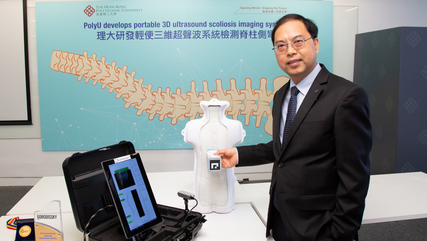
Scoliosis assessment device in a suitcase

Scoliosis is one of the most prevalent spinal deformity affecting adolescents. Early detection and regular check-up are crucial in effective scoliosis treatment. In view that X-ray assessment exposes patients to radiation and may pose increased risk for cancer, PolyU previously developed the Scolioscan – a radiation-free measurement system for accurate screening and monitoring of scoliosis using ultrasound imaging technology. Scolioscan has been installed in clinics in Australia, the Chinese mainland, Hong Kong, Italy, Macau and Netherlands, etc benefitting over 4,000 patients. A total of 23 patents for the related technology have been awarded to, or filed by, PolyU and the collaborating company.
Based on the Scolioscan technology, Prof. Zheng Yong-ping of the Department of Biomedical Engineering led a team to further develop the “Scolioscan Air” portable system, bringing the benefits of convenient and safe mass screening of scoliosis.
1. What are the advantages of Scolioscan Air?
Weighing only 5kg, the palm-sized Scolioscan Air can facilitate school-based scoliosis mass screening for youngsters anytime, anywhere. Furthermore, the technological compatibility makes Scolioscan Air readily available for commercialisation for popular use. With such small size and lightweight, the device can be fitted in a suitcase.
2. What are the special features of Scolioscan Air?
This award-winning device consists of three hardware components: i) a palm-sized wireless ultrasound probe with an optical marker mounted at its bottom; ii) a depth camera; iii) a laptop or tablet computer with dedicated software. The compact optical marker and depth camera in Scolioscan Air replace the spatial sensor used in Scolioscan. This has dramatically downsized the device whilst the high accuracy in optical spatial tracking is comparable to Scolioscan.
3. What are the latest technology developments associated with Scolioscan?
Developed by the research team, the technologies for 3D ultrasound image reconstruction, visualisation and measurement, including a fully automatic curvature measurement method and 3D spinal deformity analysis software, can be applied to Scolioscan Air. All these facilitate the device to obtain patients’ images in various postures, provide vertebra rotation and muscle-related information, and form 3D spinal models for 3D analysis of spine deformity. These technologies can also be applied to conducting prognosis and monitoring treatment outcomes, so as to establish a personalised treatment plan for individual scoliosis patient.
With funding support from the Research Impact Fund under the Research Grants Council, we are optimising our 3D ultrasound imaging system to automate the process of creating 3D spinal model. We hope that the new software can help to predict spinal curve progression, and facilitate the design of orthosis and exercises to treat scoliosis.

