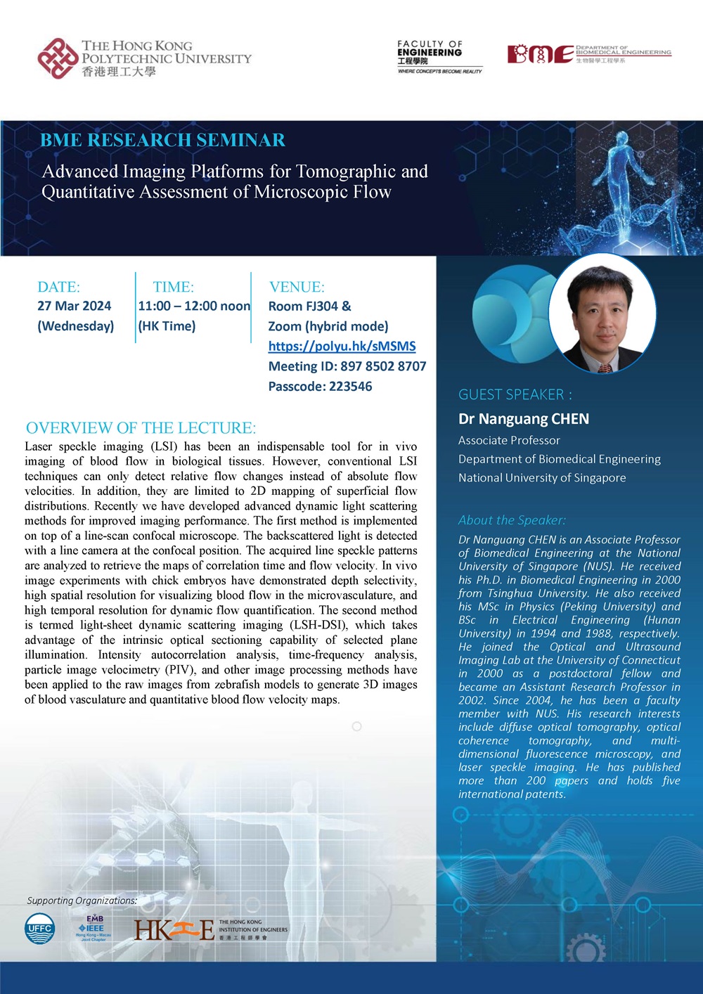
BME Research Seminar: Advanced Imaging Platforms for Tomographic and Quantitative Assessment of Microscopic Flow
Conference / Lecture
-
Date
27 Mar 2024
-
Organiser
Department of Biomedical Engineering
-
Time
11:00 - 12:00
-
Venue
FJ304 Map
Summary
BME Research Seminar: Advanced Imaging Platforms for Tomographic and Quantitative Assessment of Microscopic Flow
Zoom: https://polyu.hk/sMSMS (Meeting ID: 897 8502 8707; Passcode: 223546)
Overview of the Lecture
Laser speckle imaging (LSI) has been an indispensable tool for in vivo imaging of blood flow in biological tissues. However, conventional LSI techniques can only detect relative flow changes instead of absolute flow velocities. In addition, they are limited to 2D mapping of superficial flow distributions. Recently we have developed advanced dynamic light scattering methods for improved imaging performance. The first method is implemented on top of a line-scan confocal microscope. The backscattered light is detected with a line camera at the confocal position. The acquired line speckle patterns are analyzed to retrieve the maps of correlation time and flow velocity. In vivo image experiments with chick embryos have demonstrated depth selectivity, high spatial resolution for visualizing blood flow in the microvasculature, and high temporal resolution for dynamic flow quantification. The second method is termed light-sheet dynamic scattering imaging (LSH-DSI), which takes advantage of the intrinsic optical sectioning capability of selected plane illumination. Intensity autocorrelation analysis, time-frequency analysis, particle image velocimetry (PIV), and other image processing methods have been applied to the raw images from zebrafish models to generate 3D images of blood vasculature and quantitative blood flow velocity maps.
About the Speaker
Dr Nanguang CHEN is an Associate Professor of Biomedical Engineering at the National University of Singapore (NUS). He received his Ph.D. in Biomedical Engineering in 2000 from Tsinghua University. He also received his MSc in Physics (Peking University) and BSc in Electrical Engineering (Hunan University) in 1994 and 1988, respectively. He joined the Optical and Ultrasound Imaging Lab at the University of Connecticut in 2000 as a postdoctoral fellow and became an Assistant Research Professor in 2002. Since 2004, he has been a faculty member with NUS. His research interests include diffuse optical tomography, optical coherence tomography, and multi-dimensional fluorescence microscopy, and laser speckle imaging. He has published more than 200 papers and holds five international patents.


