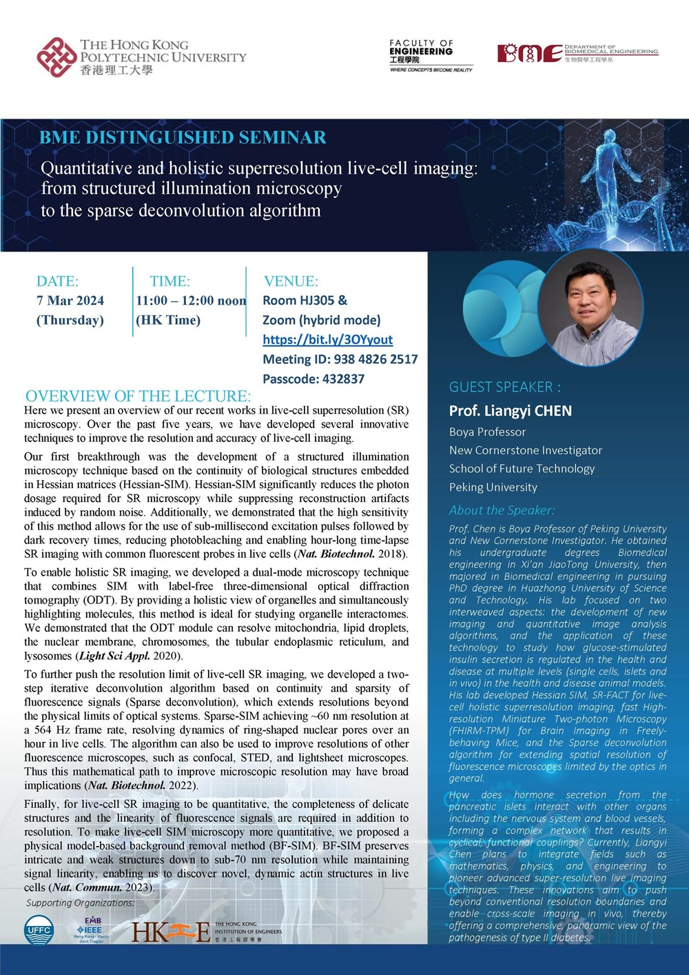
BME Research Seminar: Quantitative and Holistic Superresolution Live-cell Imaging: From Structured Illumination Microscopy to the Sparse Deconvolution Algorithm
Conference / Lecture
-
Date
07 Mar 2024
-
Organiser
Department of Biomedical Engineering
-
Time
11:00 - 12:00
-
Venue
HJ305 Map
Summary
BME Research Seminar: Quantitative and Holistic Superresolution Live-cell Imaging: From Structured Illumination Microscopy to the Sparse Deconvolution Algorithm
Zoom: https://bit.ly/3OYyout (Meeting ID: 938 4826 2517; Passcode: 432837)
Overview of the Lecture
Here we present an overview of our recent works in live-cell superresolution (SR) microscopy. Over the past five years, we have developed several innovative techniques to improve the resolution and accuracy of live-cell imaging.
Our first breakthrough was the development of a structured illumination microscopy technique based on the continuity of biological structures embedded in Hessian matrices (Hessian-SIM). Hessian-SIM significantly reduces the photon dosage required for SR microscopy while suppressing reconstruction artifacts induced by random noise. Additionally, we demonstrated that the high sensitivity of this method allows for the use of sub-millisecond excitation pulses followed by dark recovery times, reducing photobleaching and enabling hour-long time-lapse SR imaging with common fluorescent probes in live cells (Nat. Biotechnol. 2018).
To enable holistic SR imaging, we developed a dual-mode microscopy technique that combines SIM with label-free three-dimensional optical diffraction tomography (ODT). By providing a holistic view of organelles and simultaneously highlighting molecules, this method is ideal for studying organelle interactomes. We demonstrated that the ODT module can resolve mitochondria, lipid droplets, the nuclear membrane, chromosomes, the tubular endoplasmic reticulum, and lysosomes (Light Sci Appl. 2020).
To further push the resolution limit of live-cell SR imaging, we developed a two-step iterative deconvolution algorithm based on continuity and sparsity of fluorescence signals (Sparse deconvolution), which extends resolutions beyond the physical limits of optical systems. Sparse-SIM achieving ~60 nm resolution at a 564 Hz frame rate, resolving dynamics of ring-shaped nuclear pores over an hour in live cells. The algorithm can also be used to improve resolutions of other fluorescence microscopes, such as confocal, STED, and lightsheet microscopes. Thus this mathematical path to improve microscopic resolution may have broad implications (Nat. Biotechnol. 2022).
Finally, for live-cell SR imaging to be quantitative, the completeness of delicate structures and the linearity of fluorescence signals are required in addition to resolution. To make live-cell SIM microscopy more quantitative, we proposed a physical model-based background removal method (BF-SIM). BF-SIM preserves intricate and weak structures down to sub-70 nm resolution while maintaining signal linearity, enabling us to discover novel, dynamic actin structures in live cells (Nat. Commun. 2023).
About the Speaker
Prof. Chen is Boya Professor of Peking University and New Cornerstone Investigator. He obtained his undergraduate degrees Biomedical engineering in Xi’an JiaoTong University, then majored in Biomedical engineering in pursuing PhD degree in Huazhong University of Science and Technology. His lab focused on two interweaved aspects: the development of new imaging and quantitative image analysis algorithms, and the application of these technology to study how glucose-stimulated insulin secretion is regulated in the health and disease at multiple levels (single cells, islets and in vivo) in the health and disease animal models. His lab developed Hessian SIM, SR-FACT for live-cell holistic superresolution imaging, fast High-resolution Miniature Two-photon Microscopy (FHIRM-TPM) for Brain Imaging in Freely-behaving Mice, and the Sparse deconvolution algorithm for extending spatial resolution of fluorescence microscopes limited by the optics in general.
How does hormone secretion from the pancreatic islets interact with other organs including the nervous system and blood vessels, forming a complex network that results in cyclical, functional couplings? Currently, Liangyi Chen plans to integrate fields such as mathematics, physics, and engineering to pioneer advanced super-resolution live imaging techniques. These innovations aim to push beyond conventional resolution boundaries and enable cross-scale imaging in vivo, thereby offering a comprehensive, panoramic view of the pathogenesis of type II diabetes.


