
Nikon N-SIM/N-STORM/A1 Super-resolution/Confocal Microscope
Specifications:
- Standalone or correlative confocal, SIM and STORM imaging
- Eclipse Ti fully motorised inverted microscope body
- Adaptive auto-focusing system (Nikon Perfect Focus System)
- High-precision motorised stage with encoder
- Tile scanning and image stitching to produce high-resolution overview image of large tissue sections
- Motorised condenser, objective and fluorescence filter turrets
- Transmitted light detector for bright-field and differential interference contrast (DIC) imaging
- Multi-wavelength LED fibre optic illumination system (CoolLED pE-300white) with spectral coverage from ultraviolet to red region
- Environmental chamber with temperature and CO2 concentration control (Tokai Hit INU series)
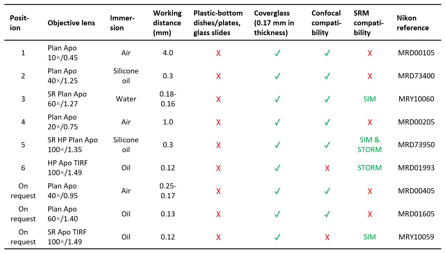
- Objectives (details):
- CFI Plan Apo 10×/0.45 Dry
- CFI Plan Apo 20×/0.75 Dry
- CFI Plan Apo 40×/0.95 Dry
- CFI Plan Apo 40×/1.25 Silicone Oil
- CFI Plan Apo 60×/1.40 Oil
- CFI SR Plan Apo 60×/1.27 Water (compatible with SIM)
- CFI SR Apo TIRF 100×/1.49 Oil (compatible with SIM)
- CFI HP Apo TIRF 100×/1.49 Oil (compatible with STORM)
- CFI SR HP Plan Apo 100×/1.35 Silicone Oil (compatible with SIM & STORM)
Usage rate
- Internal users: $215
- Collaborators: $430
- External users: $1075
Location
Y708b
Staff-in-charge
- Z215
- 34008923
- shuqi.zhang@polyu.edu.hk
- Z215
- 34008985
- idyht.ho@polyu.edu.hk