Upconversion nanoparticles are used as optical probes in fluorescence resonance energy transfer
assay for ultrasensitive flu virus detection.
Overcoming traditional limitations
|
This method converts low-energy photons into high-energy photons, or more specifically, invisible near infrared photons into visible green photons. Dr Yang Mo |
 |
Prof. Hao explained that the work on the biosensor had developed out of collaboration of the two teams undertook in 2014 on the use of upconversion nanoparticles (UCNPs) as optical probes in fluorescence resonance energy transfer assay for ultrasensitive flu virus detection. Concerned at the time that the H7N9 flu virus posed a threat to public safety and animal life, the teams knew that the traditional optical luminescence detection is limited by the use of high energy light sources and a low limit of detection.
When working on the new sensor, the teams were still seeking to overcome the limits of traditional biological methods. The two main methods traditionally used in flu virus detection are reverse transcription-polymerase chain reaction (RT-PCR) and enzyme-linked immunosorbent assay (ELISA). RT-PCR involves changing the Ribonucleic acid (RNA) of interest – which carries genetic information in viruses such as influenza and Ebola – to DNA, which is then duplicated using a polymerase chain reaction in a thermocycler until its levels can be detected in a very costly and labour-intensive process. ELISA, while less costly as it involves the attraction between the antigen of the target and its antibody to produce colour change on a plastic well plate, has relatively low sensitivity and requires high-quality samples. Both methods are time consuming and of limited value as frontline, on-site diagnostic tools.
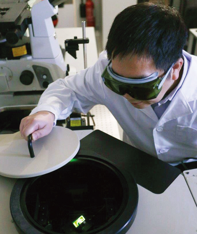
Researcher analyses the sensitivity of the nanobiosensor.
Optical detection, which has high sensitivity, is thus a more viable option for rapid virus detection. Yet the conventional method used in biological application, downconversion luminescence, is excited by high-energy sources such as ultraviolet light, which can damage genetic material and induce background florescence. The teams thus focused on upconversion luminescence. Dr Yang explained, “this method converts lowenergy photons into high-energy photons, or more specifically, invisible near infrared photons into visible green photons”.
The overall technique again relies on UCNPs and is akin to matching two magnets with their force of attraction. UCNPs are conjugated with a probe oligo (a short DNA strand otherwise known as an oligonucleotide), the base pairs of which are complementary with that of a gold nanoparticle (AuNP) flu virus oligo. Given the complementary nature of the base pairs of the two oligos, they work like positive and negative magnetic poles being drawn together in a process called oligo hybridisation. Dr Yang further explained, “when illuminated by a portable near-infrared laser pen, the UCNPs emit visible green light and the AuNPs absorb the light. The concentration of the targeted flu virus can then be easily quantified by measuring the decrease in green light intensity.” This, put simply, is detectable by the naked eye.
Unlike the traditional methods, this novel method is a simple procedure that requires no expensive machinery or sophisticated operating skills. Moreover, in comparison to the conventional downconversion luminescent technique, it causes little damage to genetic material and induces no background fluorescence. Just as importantly, it can be modified through the design of a complementary probe once the genetic sequence of a newly targeted virus is known, opening the possibility of detecting other types of viruses.
